0.0 Bachelorarbeit - Gesamtübersicht
0.2 Abstract
Abstract Text
Link zum Original
1.0 Introduction
1.1. From Wernicke’s to Dual Stream
1.1.1 Two Streams Hypothesis
The brain’s ability of efficiently processing input relies on a sophisticated methods - separating streams that compute different aspects of the inputs. One that computes information about the object itself - shape, colour, type - and the other streams computes motion and spatial information - where is the object in relation to oneself, where is it moving towards?
The discovery of two streams in the brain of the dorsal where and the ventral what stream already occurred in the 1980s, especially for visual inputs. They form a clear pattern starting from the visual cortex and moving anteriorly into the prefrontal cortex. (Quelle?)
The dorsal stream passes through the parietal cortex and terminates in the prefrontal cortex. From there top down attention is directed by the FEF.
The ventral visual stream passes down the temporal lobe passing by FFA and other recognition areas ending in prefrontal cortex. From there top down attention is directed by IFJ (Bedini & Baldauf (2021))For clarity in this paper we will use “where”-stream for the posterodorsal stream and “what” for the anteroventral stream due to some areas that could be location-wise assinged to a different stream than its acutal functional connectivity. Therefore we focus on connectivity, because this is more significant for attention than its pure location.
1.1.2 Dual processing in the auditory cortex
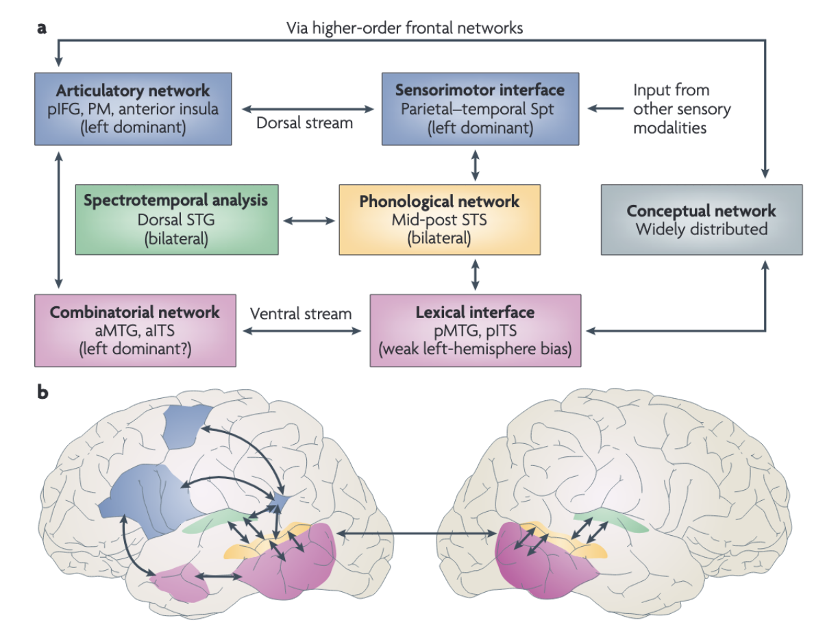
Historically, the neural basis of language comprehension was thought to be localized in a single region: Wernicke’s area predominantly in the left superior temporal gyrus (STG). This classic model dominanted neuroscience for over a century. However, this monolithic was challenged in the 1970s and 1980s by studies revealing that lesions to the left STG did not necessarily lead to deficits in the auditory comprehension, but rather caused deficits in speech production (Hickok & Poeppel 2007 - Nature). These findings lead to a fundamental re-evaluation of auditory cortical organization.
Today a similar structure is widly accepted for auditory inputs as well. What needs to be researched is whether FEF and IFJ function is attention hubs also for the auditory pathways and not only for visual. This is this research looking into.
The auditory system is commonly described as consisting of two major processing streams similar to the visual system with mainly two pathways. The posterodorsal where-stream and the anteriorventral what-stream.
State of the art in the pathway research of auditory cortex are especially developed by Hickok & Poeppel 2007 - Nature and Rolls et al. (2023) - Cerebral Cortex.⇒ soll ich das hier noch weiter ausführen?
also die figure erklären?
wie weit soll ich hier gehen?
Figure 1: Hickock and Poeppel developed a framework explaining the dorsal and ventral auditory streams connecting them to higher-order frontal networks.
Link zum Original1.2 The Gap, Top-Down Control of Auditory Streams
The division into dorsal and ventral pathways is well-documented(Ahveninen et al. (2006) - PNAS, Hickok & Poeppel 2007 - Nature), but misses how these streams are top-down coordinated. To manage this, the top-down attention is required to filter relevant information and suppress distractors (De Vries & Baldauf (2021) - Journal of Neuroscience). The question remains: Which prefrontal regions act as the conductors for these auditory streams?
In the visual system this question has been answered. Recent neuroimagning research has identified the double-association in the prefrontal cortex, conducted by the FEF and IFJ:
- Frontal Eye Field (FEF): a dorsal region specialized for spatial processing and oculomotor control. It performs a strong top-down influence over the visual where (“dorsal”) stream (Bedini & Baldauf (2021), Bedini (2023) - Brain Structure).
- anterior Inferior Frontal Junction (IFJa): a region located ventrally from the FEF and controls the visual wha “ventral” stream specializing on object-based attention (Bedini & Baldauf (2021))
This is demonstrates a clear “control hub” hierarchy for vision. For the auditory system, the exact top-down “control hubs” are less defined. While Rolls et al. (2023) - Cerebral Cortex and Frühholz (2015) - NeuroImage point out especially IFG and Broca’s areas, such as BA44, 44, BA45, 45, as hubs, this is mainly for the semantic language pathway. There is still a piece missing for the exact controllers, especially for the auditory where-stream. Hickok & Poeppel 2007 - Nature mention a dorsal-motor pathway which might be located in the PT (Hickok & Poeppel 2007 - Nature).
Link zum OriginalLink zum Original1.3 Hypothesis, A supramodal organization
We hypothesize that the functional connections of the auditory what and where streams are supramodal within the prefrontal cortex both being coordinated through top-down attention mechanisms from the FEF and IFJ.
In detail:
- The seeds of the Frontal Eye Field (FEF) should preferentially connect to the auditory where-stream
- The seeds of the Inferior Frontal Junction (IFJ) should preferentially connect to the auditory what-stream
Because both FEF and IFJ are known to direct top-down attentional control in the visual domain, we propose that they may play an analogous role for auditory attention as well along the where and what pathways.
In summary we test whether the prefrontal cortex shows the same division of attention direction in the auditory stream as is found in the visual stream.
Link zum Original
2.0 Theoretical Background
Transclude of 2.1-The-Auditory-What-("Ventral")-Stream#21-the-auditory-what-ventral-streamTransclude of 2.2-The-Auditory-Where-("Dorsal")-Stream#22-the-auditory-where-dorsal-stream2.3 Top Down Control
1. Subheading
Hier schreiben…
2. Subheading
Link zum Original
Link zum Original2.4 Anatomical and Classification Challenges
1. Subheading
Hier schreiben…
2. Subheading
Link zum Original
3.0 Methods
Outline 3.1 Data Acquisition & Preprocessing
Link zum Original
- Core Argument dieses Kapitels definieren
3.2 Cortical Parcellation with The HCP-MMP1 Atlas
1. Subheading
Hier schreiben…
2. Subheading
Link zum Original3.3 Selection of Regions of Interest (ROIs)
To test the hypothesis of supramodal prefrontal control, we defined a set of Regions of Interest (ROIs) based on the multimodal parcellation of the Human Connectome Project (HCP-MMP1) by Glasser et al. (2016) - Nature. This atlas maps cortical areas based on a combination of cortical architecture (myelin), function (task-fMRI) and connectivity, providing a significantly higher precision than traditional Brodmann maps. ROIs were divided into three categories: (1) Prefrontal Seed Regions (the controllers), (2) Auditory Where-Stream targets (“dorsal/spatial”), (3) Auditory What-Stream targets (“Ventral/objects”).
The selection of ROIs is based on previous research, mainly by Rolls et al. (2023) - Cerebral Cortex about connectivity of auditory areas using the Glasser Altas Glasser et al. (2016) - Nature
In general the amount of ROIs found in both streams is far from balanced. There are more areas assigned to the what/ventral auditory stream. This makes sense since language comprehension requires more cotrical compute than locating sounds.
3.3.1 Prefrontal Seed Regions
We selected distict prefrontal “control hubs” as seed regions based on the functional dissociation described by Bedini & Baldauf (2021) in the visual stream.
- Frontal Eye Field (FEF): We defined the FEF using the specific Glasser label FEF. This region is located in the caudal middle frontal gyrus, ventral to the junction of the superior sulcus (sPCS) and superior frontal sulcus (SFS).
- The FEF contains topographic maps of contralateral space and is a core node of the Dorsal Attention Network (DAN), responsible for spatial attentional and oculomotor control.
- anterior Inferior Frontal Junction (IFJa): We defined the IFJa using the specific Glasser label IFJa. This region is located at the junction of the inferior precentral sulcus (iPCS) and the inferior frontal sulcus (IFS).
- Unlike the FEF, the IFJa is part of the Frontoparietal Network (FPN) and shows multiple-demand characteristics.
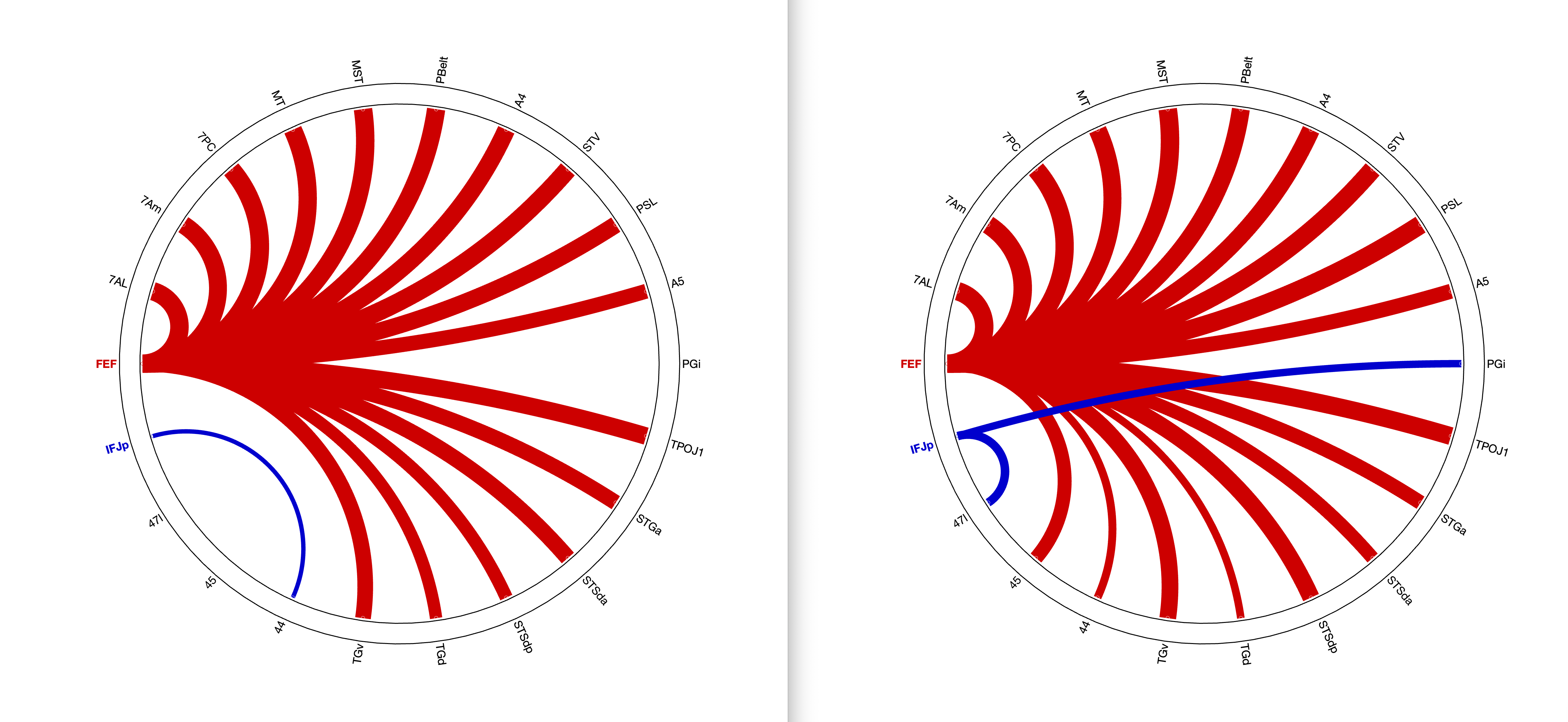
IFJa vs IFJp
Most papers soeak of the IFJ as a whole unit, but Bedini (2023) - Brain Structure found that IFJa and IFJp exhibit totally different connectivity patterns.
While the majority of neuroimaging literature treats the IFJ as a monolithic unit, recent evidence suggests a functional and conenctional dossiociation between its anterior (IFJa) and posterior (IFJp) subdivisions. We utilized the high resolution of the HCP-MMP1 to make use of this distinction.
According to Bedini & Baldauf (2021), IFJp is a core node of tthe multiple demand system and the FPN. It is activated by tasks involving reasoning and math tasks, serving as a general-purpose executive mediator
IFJp: According to the IFJp belongs to the multiple demand system and might serve as a mediator for those areas.
IFJa: functions as a mediator for lower-order regions especially for language related regions Bedini & Baldauf (2021)
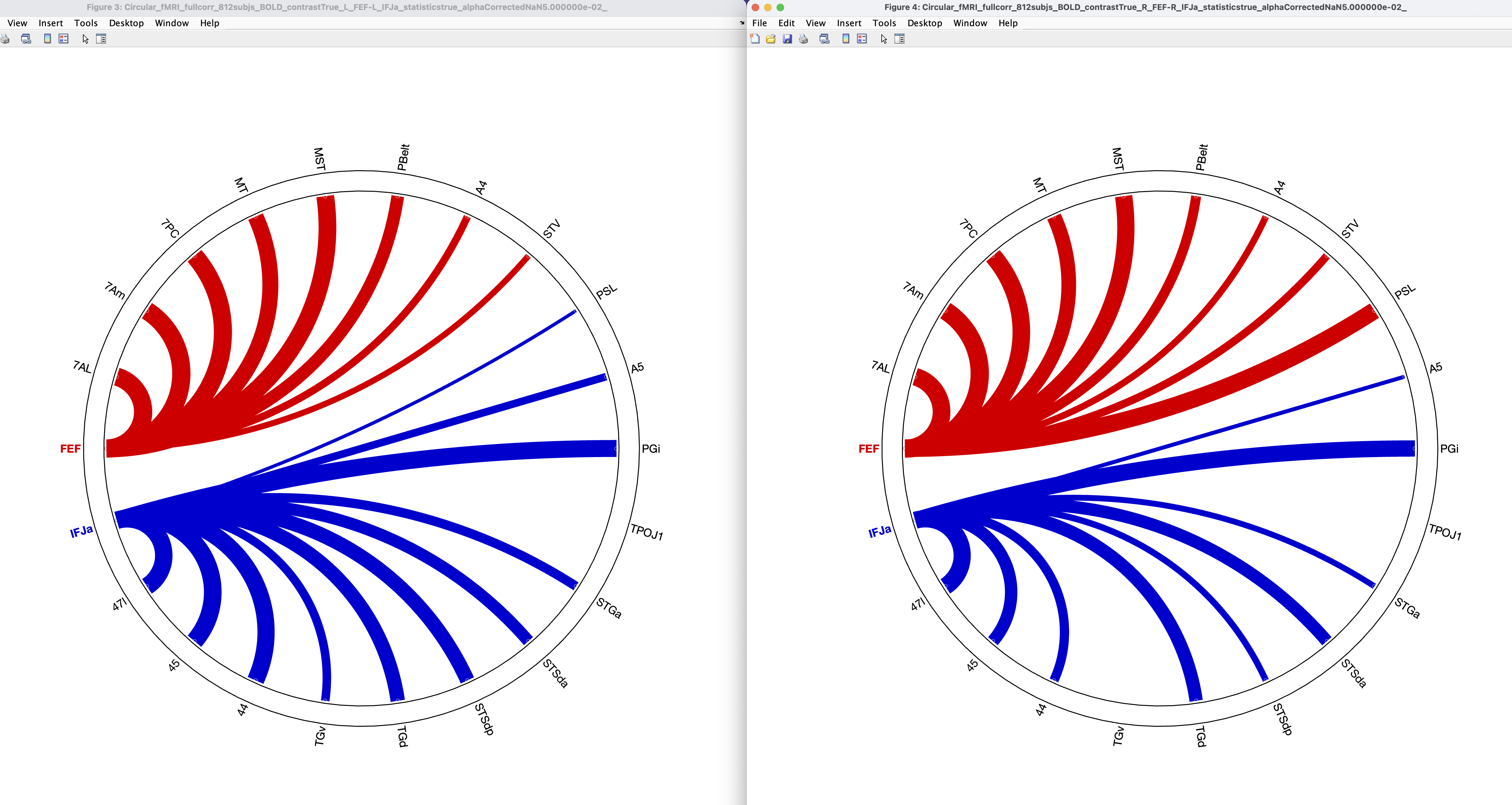
This is why we contrasted IFJa and IFJp and focus on IFJa as a seed region for the auditory what-pathway. In figure … we contrasted functional connectivity of both the IFJa and IFJp.
3.3.2 Target Definitions
3.3.2.1 ROIs for the What-Stream
Since the what-stream mainly focusses on semantics and comprehension, it is the main speech processing pathway - as in the visual stream for object recognition.
There is a lot of research on itStarting from the core and belt regions and moving anteriorly to the IFJ, we found following regions belonging certainly to the what-stream:
STGa (aSTG old description) is according to Ahveninen et al. (2006) - PNAS and the connectivity to TA2 from Glasser et al. (2016) - Nature certainly part of the what-stream. STGa has also connectivity to IFG. Also a bit of dorsal stream to IFG and fOP but weaker.
3.3.2.1 ROIs for the Where-Stream
The research on the auditory where-stream is a bit thin. Mainly I focussed on Rolls et al. (2023) - Cerebral Cortex for the connectivity between the areas.
Previous research shows that all STG subregions connect to the fOP via dorsal pathways, which could mean that STG generally belongs to the dorsal stream.
BUT3.3.2.1.1 FOP
fOP areas are vage. the only source I found was Frühholz (2015) - NeuroImage in which it says that BA44, 44 is part of fOP and therefore it could be part of the dorsal pathway.
FOP1 (Frontal Opercular Area 1): Located in the frontal operculum. According to Frühholz (2015) - NeuroImage, the fOP is a primary target of the auditory dorsal pathway (Frühholz (2015) - NeuroImage). Rolls et al. (2023) - Cerebral Cortex associated fOP regions with the “Group 3” network (including A4, A5, MT), linking it to somatosensory and motion processing. Inclusion of FOP1 captures the frontal endpoint of the “Sound to Motor” stream described by Hickok & Poeppel (2004) - Cognition, Hickok & Poeppel 2007 - Nature.
3.3.2.1.2 Spatial supporting regions 7AL, 7AM and 7PC
According to Rolls et al. (2023) - Cerebral Cortex, the areas 7AL, 7Am, 7PC could all be involved in spatial processing within the auditory stream. Since we hypothesize supramodality in which the FEF directs visual as well as auditory signals, it is plausible that letter named regions also play a significant role in spatial processing of auditory tasks.
Though, one could argue MT and MST also play a role in motion, but as studies show, there is no response of MT/MST for auditory stimuli only ⇒ Study raussuchen, da gab es eine!! Siehe Source
3.3.2.1.3 PSL
According to Rolls et al. (2023) - Cerebral Cortex PSL is involved in language-semantic functions, but Dureux (2024) shows that PSL is unresponsive to auditive stimuli (Dureux (2024)) suggesting PSL could be a highly specialized area.
It has high conenctivity to STS along with TPOJ1, STV, PSL, TGv, TGd, PGi which could suggest PSL being part of the Auditory What-Stream (Ventral).
Link zum Original3.4 Statistical Analysis
3.4.1 Functional Connectivity
This study utilizes functional connectivity (FC) as the statistical dependency of BOLD-signal-time stamps (Bloof Oxygen Level Dependent) defined in spatially divided cortical regions (Friston (1994)).
- for each subject the middle time slots of the seed regions (FEF, IFJa) as well as the auditory target areas are extracted.
- the study uses pearson-correlation coefficient as a primary measurement for functional coupling
- using it for resting-state fMRI (Biswal (1995))
3.4.2 Partial vs. Full Correlation
When comparing brain regions (or variables in general) it is essential to differentiate between the following for the interpretation of networks:
Full Correlation: Calculates the relationship between A and B without taking other variables into account. If A and B both correlate strongly with C, full correlation shows a connection between A and B, which might not even exist (indirect correlation)
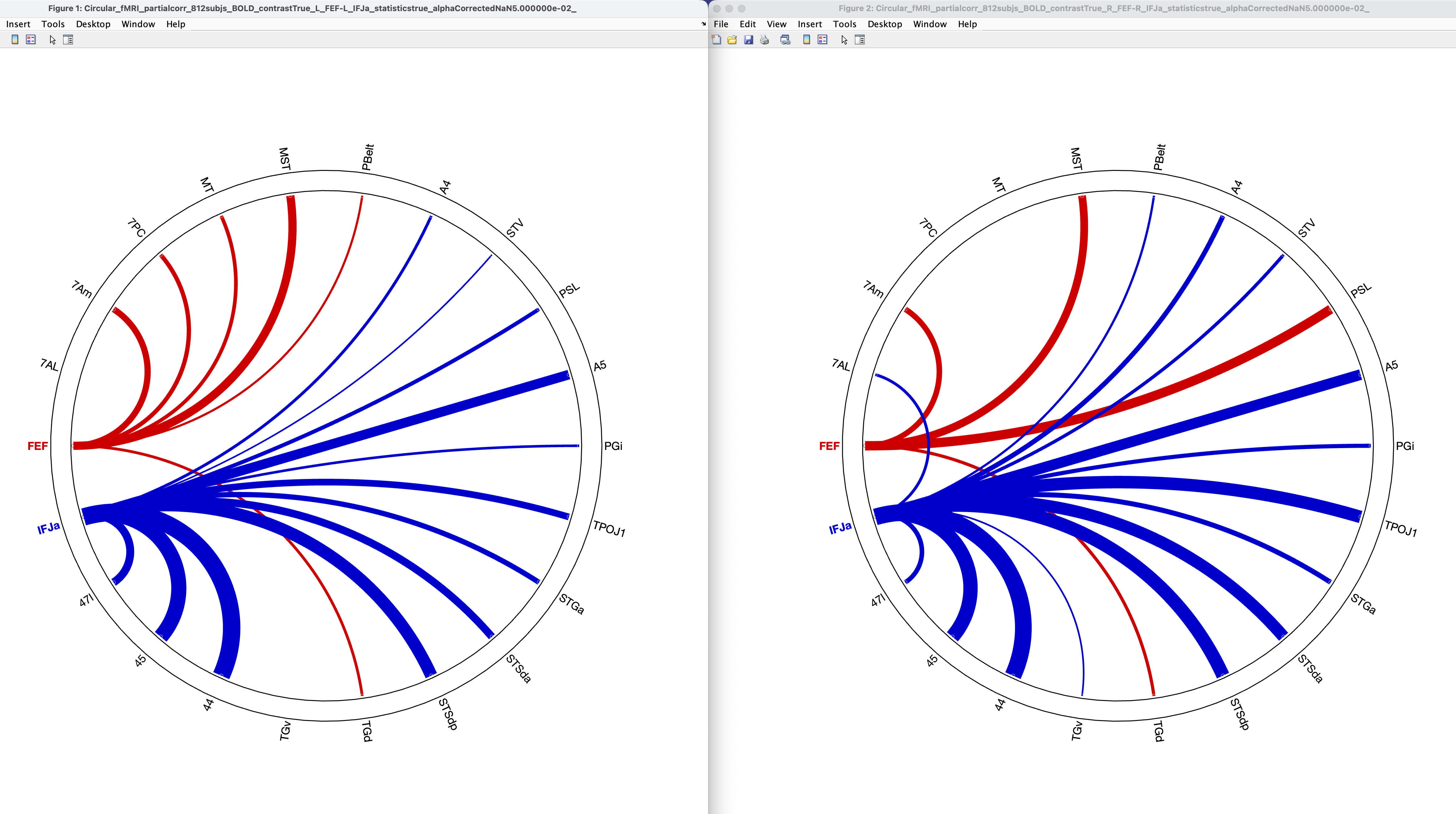
Partial Correlation: Calculates the realtionship betweenA and B after subtracting the infuence of other variables. Hence, it shows functional connectivity without the noise of other brain areas being involved. (Marrelec 2006 - NeuroImage) (Smith (2011))
In this paper, we use both full correlation for the global picture and partial correlation for the remaining connections after filtering out the other regions, whichis robust to indirect influences.
3.4.4 Correction for Multiple Tests (FDR)
The risk of more positive results due to many correlation analyses while computing several correlations for many cortical regions at the same time rises.
- Therefore we use False Discovery Rate (FDR) correction to counter this effect.
- In contrast to the rigorous Bonferroni-correction, which often turns out to be very conservative and might cover real effects, FDR correction offers a higher statistical power through checking the expected amount of rejected null hypotheses.
- in all connecitivty results there is FDR corrrection for multiple comparisons, while we used the signifance of q < 0,05 (FDR-corrected p).
Quelle: Benjamini, Y., & Hochberg, Y. (1995)
Link zum Original3.5 Brain Behaviour Correlation
1. Subheading
Hier schreiben…
2. Subheading
Link zum Original
Link zum Original
4.0 Results
4.1 Global Connectivity Patterns
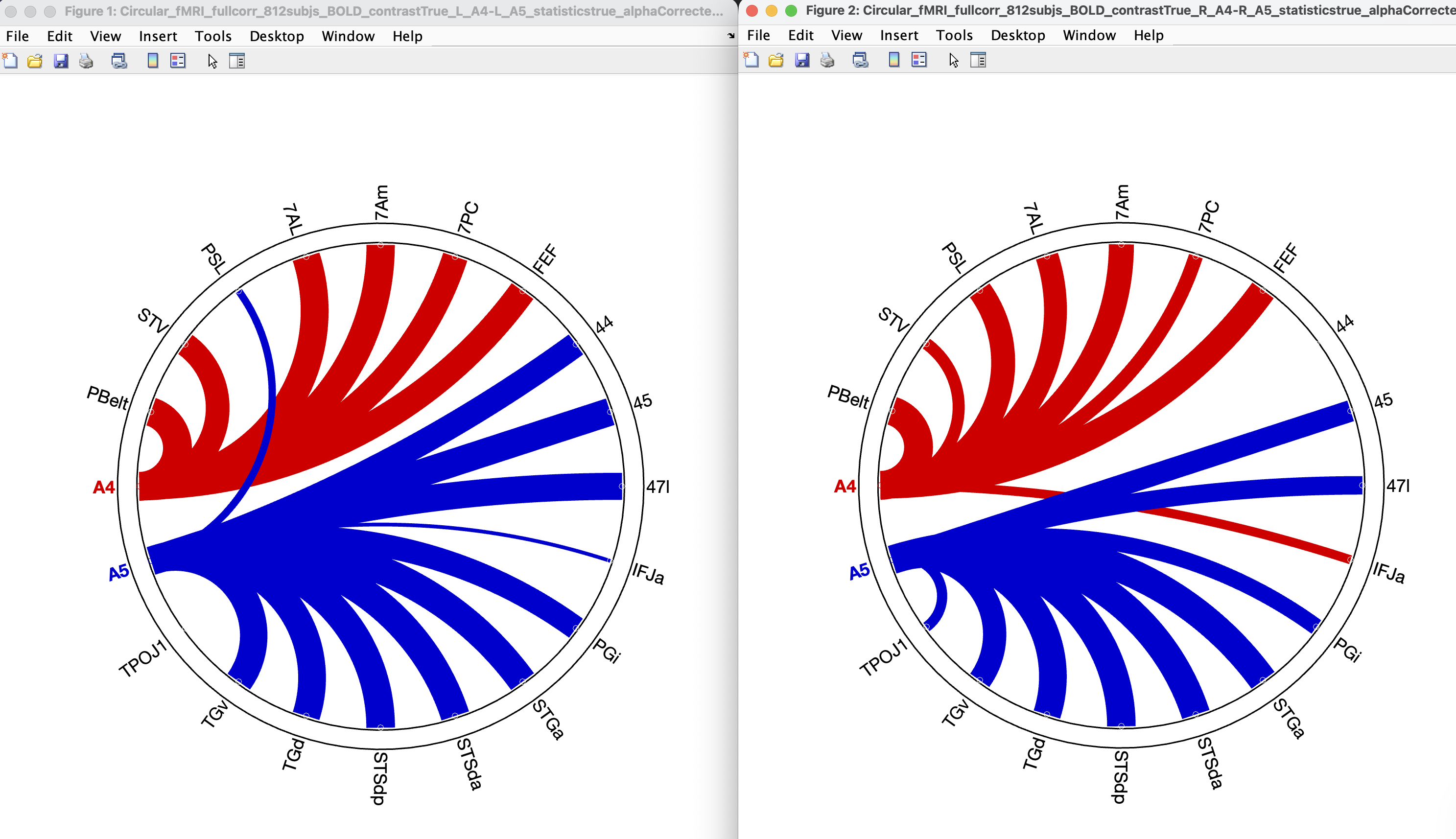
4.1.1 FEF vs. IFJa connectivity
In the analysis we compared the two seed regions FEF and IFJa in both hemispheres. Looking at functional connectivity with full correlation we found high correlation of FEF to spatial auditory areas according to Rolls et al. (2023) - Cerebral Cortex where 7AL, 7Am, 7PC might play a role in spatial orientation within the auditory where-stream. This connectivity might be running via the SLF2 (Superior Longitudinal Fasculus), partly SLF3 (Bedini (n.d.)), which mediates spatial-motor and auditory communication.
First if we look at functional connectivity we see a clear pattern that connects the FEF to the Where-pathway and auditory areas.
Left vs right hemisphere, full connectivity
Als Null-Seed, der beweist, dass es wirklich um IFJa geht und im posterior part nichts passiert.
FEF vs IFJp:
Link zum Original
Transclude of 4.2-Testing-the-"Where"-Stream-(FEF-Connectivity)#42-testing-the-where-stream-fef-connectivityTransclude of 4.3-Testing-the-"What"-Stream-(IFJa-Connectivity)#43-testing-the-what-stream-ifja-connectivityLink zum Original4.4 Resolving Ambiguities
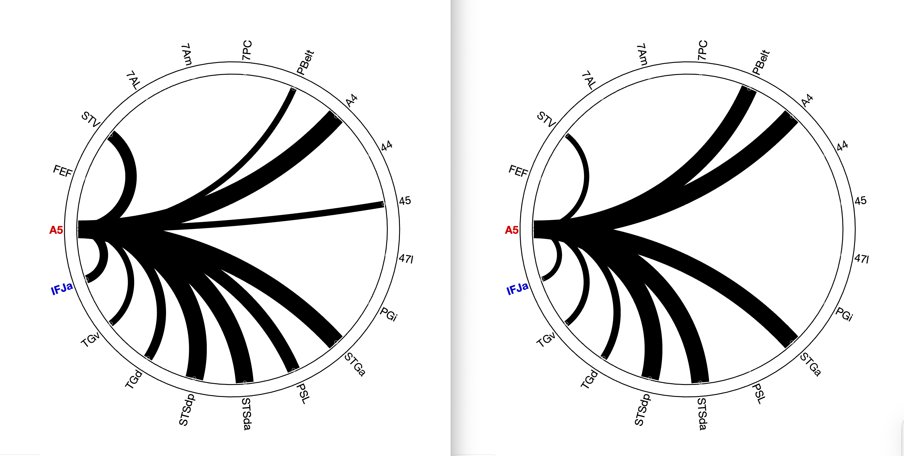
A4 and A5
anatomic locations of A4 and A5 are in the parabelt cortex and are in the posterior part of the superior temporal gyrus (STG). They form the transition from early auditory cortex to specialized streams. In luterature they are being reffered as to the starting point of the dorsal stream (Quelle benötigt). Nach Rolls ist es unsicher, ob A5 zu Where oder zum What pathway gehört, da A5 sowohl connectivity zu Area 44 und IFJa zeigt, als auch zu MT und MST, welche klar zum where-pathway des visuellen streams gehören.
A4
In Glasser et al. (2016) - Nature A4 is defined as functionally different from its neighbours:
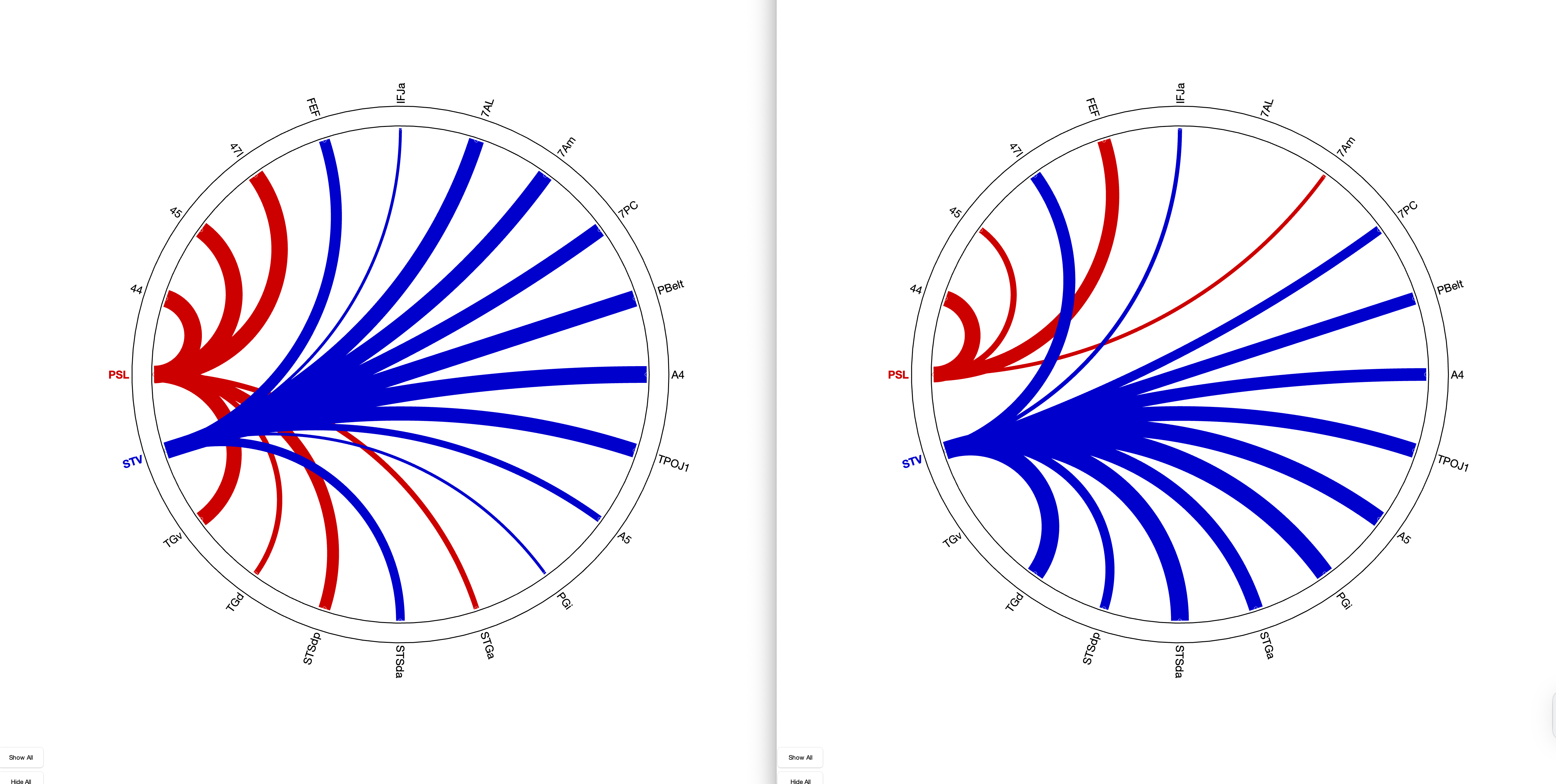
- in contrast to STV, A4 shows a weaker connectivity to visial area PCV
- also weaker connectivity to 55b than PSL
PSL the division
PSL (perisylvian Language Area) is located at the intersection of temporal and parietal lobe in the sylvian region. In the cluster of Glasser et al. (2016) - Nature PSL is classified as a semantic group (Group 3), that process auditory information and integrates it into the language system.
According to Dureux (2024) PSL is unresponsive to all tested auditive stimuli.
- unresponsive to all tested auditive stimuli according to Dureux (2024) auch
in contrast to STV, we see
STV surprise
Link zum Original
5.0 Discussion
1. Subheading
Hier schreiben…
2. Subheading
Link zum Original
6.0 References
1. Subheading
Hier schreiben…
2. Subheading
Link zum Original
7.0 Appendix
1. Subheading
Hier schreiben…
2. Subheading
Link zum Original
8.0 Declaration
1. Subheading
Hier schreiben…
2. Subheading
Link zum Original
9.0 Abbreviations
1. Subheading
Hier schreiben…
2. Subheading
Link zum Original