Frühholz (2015) - NeuroImage
- Paper gelesen?
- Infos rausgeschrieben?
Frühholz, S., Gschwind, M., & Grandjean, D. (2015). Bilateral dorsal and ventral fiber pathways for the processing of affective prosody identified by probabilistic fiber tracking. NeuroImage, 109, 27–34. https://doi.org/10.1016/j.neuroimage.2015.01.016
-
Dorsal and ventral pathways for syntacto-semantic speech processing in the left hemisphere are represented in the dual-stream model of auditory processing.
-
The results also suggest the existence of a dual-stream processing in the right hemisphere, and a general predominance of the dorsal pathways in both hemispheres underlying the neural processing of affective prosody in an extended temporo-frontal network.
-
Recent studies (Friederici et al., 2006; Rauschecker and Scott, 2009; Saur et al., 2008) have predominantly identified left hemispheric processing pathways within a dual-stream model of auditory processing (Hickok and Poeppel, 2007). They include ventral pathways from anterior superior temporal gyrus (STG) to the anterior inferior frontal gyrus (IFG) and dorsal pathways, which project to the posterior IFG via the posterior STG (Hickok and Poeppel, 2007; Rauschecker and Scott, 2009).
- bestätigt, was wir eh schon wissen
-
Especially the dorsal pathway seems strongly left lateralized (Hickok and Poeppel, 2007). The ventral pathways convey sound-invariant meaning (Belin and Zatorre, 2000b; Rauschecker and Scott, 2009), such as speech semantics (Hagoort, 2005).
-
The dorsal pathways serve sound-to-motor mapping (Saur et al., 2008) and the processing of temporal auditory sequences (Belin and Zatorre, 2000b; Rauschecker and Scott, 2009), which are also necessary for the understanding of speech syntax (Friederici et al., 2006).
-
Compared to a predominant role of the left brain for syntacto-semantic processing (Specht, 2014), the emotional intonation in speech, that is the affective prosody, strongly, but not exclusively, activates regions in right STG and IFG (e.g. Alba-Ferrara et al., 2011; Beaucousin et al., 2007; Ethofer et al., 2006; Fruhholz et al., 2012).
-
Ventral and dorsal pathways, for example, are supposed to originate in multiple STG seed regions (Friederici, 2011; Fruhholz et al., 2012). Furthermore, these pathways probably terminate in the anterior as well as in the posterior IFG (Fruhholz and Grandjean, 2013b)
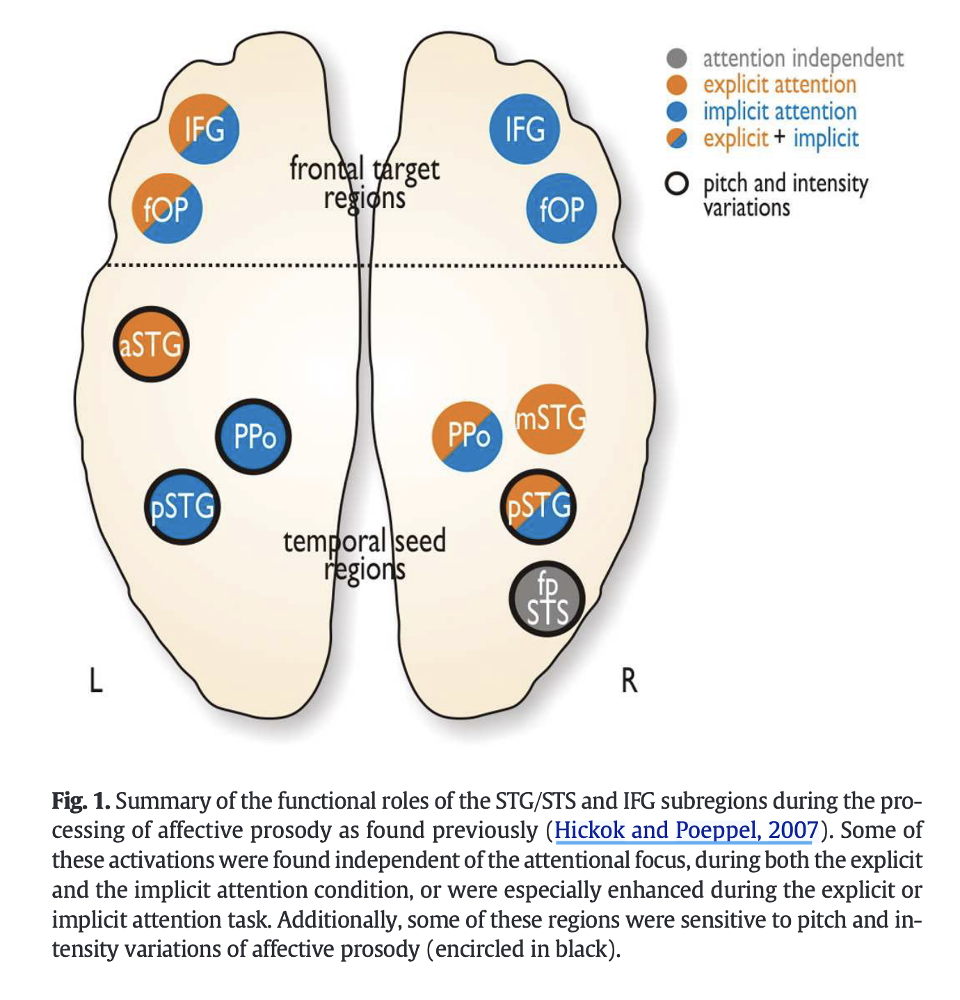
STG-Regionen:
- The right compared with left IFG showed a sensitivity to speech prosody during implicit attention, while left IFG subregions responded to affective prosody during both attentional conditions. All STG subregions showed a sensitivity to speech prosody both for the explicit and implicit attention condition, but right mid STG (mSTG) and left anterior STG (aSTG) showed stronger sensitivity during the explicit attention condition, whereby the latter regions showed a general main effect for the explicit compared with the implicit task, and thus might reflect a rather general evaluation of voices independent of the emotion. Furthermore, regions in the right posterior STG (fundus of the posterior superior temporal sulcus (fpSTS), posterior STG (pSTG)) and all left STG subregions were sensitive to the pitch and intensity variations in affective prosody, which are one of the main acoustic features of affective prosody (Banse and Scherer, 1996; Patel et al., 2011).
Und hier ventral/dorsal pathways according to Frühholz:
Results:
- The anterior STG (aSTG) was connected to the IFG via a strong ventral pathway consisting of the inferior longitudinal fasciculus (ILF) and the inferior fronto-occipital fasciculus (IFOF) in its posterior portion, and the extreme capsule (EmC) in the anterior portion (Fig. 5A), as confirmed with a standard white matter atlas
- The aSTG was also connected to the IFG via a dorsal pathway, but the aSTG–IFG connectivity displayed a higher pathway probability (PP) via the ventral compared with the dorsal pathway (t14 = 18.874, P = 2.35 × 10⁻¹¹) (Fig. 3D).
- The aSTG finally showed a connection to the left frontal operculum (fOP) by dorsal pathways. Unlike the aSTG, the left polare plane (PPo) and the posterior STG (pSTG) were connected to both the IFG and the fOP only via dorsal pathways. The strongest CP originating from the STG seed regions was actually found for the pSTG and targeting all frontal regions (main effect for the factor temporal seed: F1.44,20.16 = 7.469, P = 0.007, GreenhouseGeisser (GG) corrected), especially for the comparison of the pSTG compared with the PPo (planned posthoc comparison: P = 7.74 × 10⁻⁴).
- we found that while all subregions in the STG were connected to the right fOP via dorsal pathways, only the most posterior region in the right fundus of the posterior superior temporal sulcus (fpSTS) was connected to the right IFG via equally strong dorsal and ventral pathways (i.e. indicated by a nonsignificant effect of for the comparison of the dorsal and ventral PP; t14 = 0.023, P = 0.982) as well as to the fOP via a dorsal pathway.
- The right PPo, mid STG (mSTG), and pSTG were connected only to the fOP via dorsal pathways. The least CP was found for the mSTG, especially as compared with the pSTG and the PPo
- we revealed some left-hemispheric temporofrontal connections for affective prosody processing, especially dorsal connection between the anterior STG and the inferior frontal cortex, in addition to those described recently for auditory processing of vocalizations and speech (Ethofer et al., 2012; Glasser and Rilling, 2008; Rauschecker and Scott, 2009; Saur et al., 2008)
- we now provide quantitative description as well as evidence for the specific subregions in STC and IFC, which are structurally connected by the right ventral pathway. Second, we found an overall bilateral predominance of the dorsal pathway for processing affective prosody.
Methods used by Frühholz:
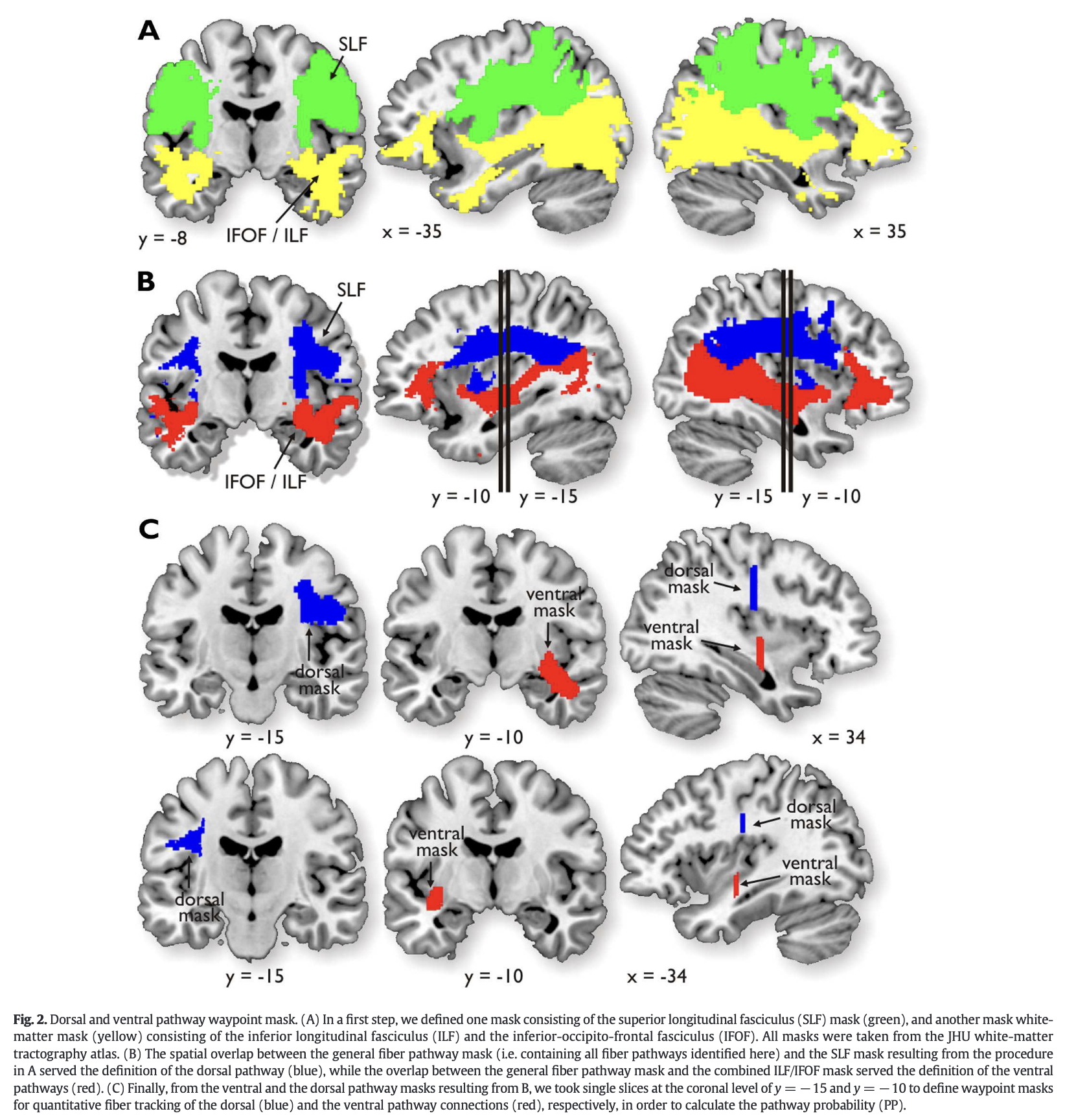
- In order to estimate the relative connectivity of the dorsal and the ventral pathways, we then conducted two additional analyses on the second stage. We included waypoint masks for the dorsal and the ventral pathway, which constrained the tracking algorithm to fibers that only passed through the respective masks (Fig. 2). These dorsal and ventral waypoint masks were generated as follows. First, we defined superior longitudinal fasciculus (SLF) masks taken from the JHU white-matter tractography atlas at a probability threshold of 100% (Hua et al., 2008). The SLF is the main dorsal longitudinal fiber bundle connecting posterior and anterior brain regions, as has been frequently reported to contain fiber connections between regions involved in auditory communication (Hua et al., 2008) (Fig. 2A). Furthermore, we defined a white-matter mask by combining the inferior longitudinal fasciculus (ILF) and the inferior fronto-occipital fasciculus (IFOF) taken from the JHU atlas, as the main ventral longitudinal fiber bundles (Friederici, 2011; Saur et al., 2008). Subsequently, we created a general white-matter pathway mask by summing up all fiber pathways from all seed and target regions across all participants. The spatial overlap between this general fiber pathway mask and the SLF mask resulting from the procedure in the first step served the definition of the dorsal pathway (Fig. 2B). The overlap between the general fiber pathway mask and the combined ILF/IFOF mask served the definition of the ventral fiber pathways.
voice-sensitive cortex:
- We took three subregions in the voice-sensitive cortex of the left hemisphere (pSTG [MNI xyz − 68 − 27 6], planum polare (PPo) [− 50 −10 4], aSTG [−56 11 − 10]), and two IFG subre- gions, one located more posterior in the frontal operculum (fOP; BA 44; [− 51 13 14]), and one more anterior in the IFG (BA 47; [− 44 29 0]) (Fig. 2B). In the right hemisphere we used the previously found four STG subregions (fpSTS [45 −34 4], pSTG [69 −22 4], mSTG [66 −3 2], PPo [53 −4 −4]), and two IFC subregions, one located more posteriorly in the fOP (BA 44; [48 13 −2], and one more anteriorly in the IFG (BA 47, [51 32 −2]) (Fig. 3B). Note that all four right STG subregions except PPo, were located in the voice-sensitive cortex, as defined by a standard voice localizer scan (see Belin and Zatorre, 2000a
see also
Tags: neuroscience science source
Superlink: 050 🧠Neuroscience
Created: 2025-11-12 22:14