Glasser, M. F., Coalson, T. S., Robinson, E. C., Hacker, C. D., Harwell, J., Yacoub, E., Ugurbil, K., Andersson, J., Beckmann, C. F., Jenkinson, M., Smith, S. M., & Van Essen, D. C. (2016). A multi-modal parcellation of human cerebral cortex. Nature, 536(7615), 171–178.
https://doi.org/10.1038/nature18933
- paper gelesen?
- Infos rausgeschrieben?
Glasser et al. (2016) - Nature
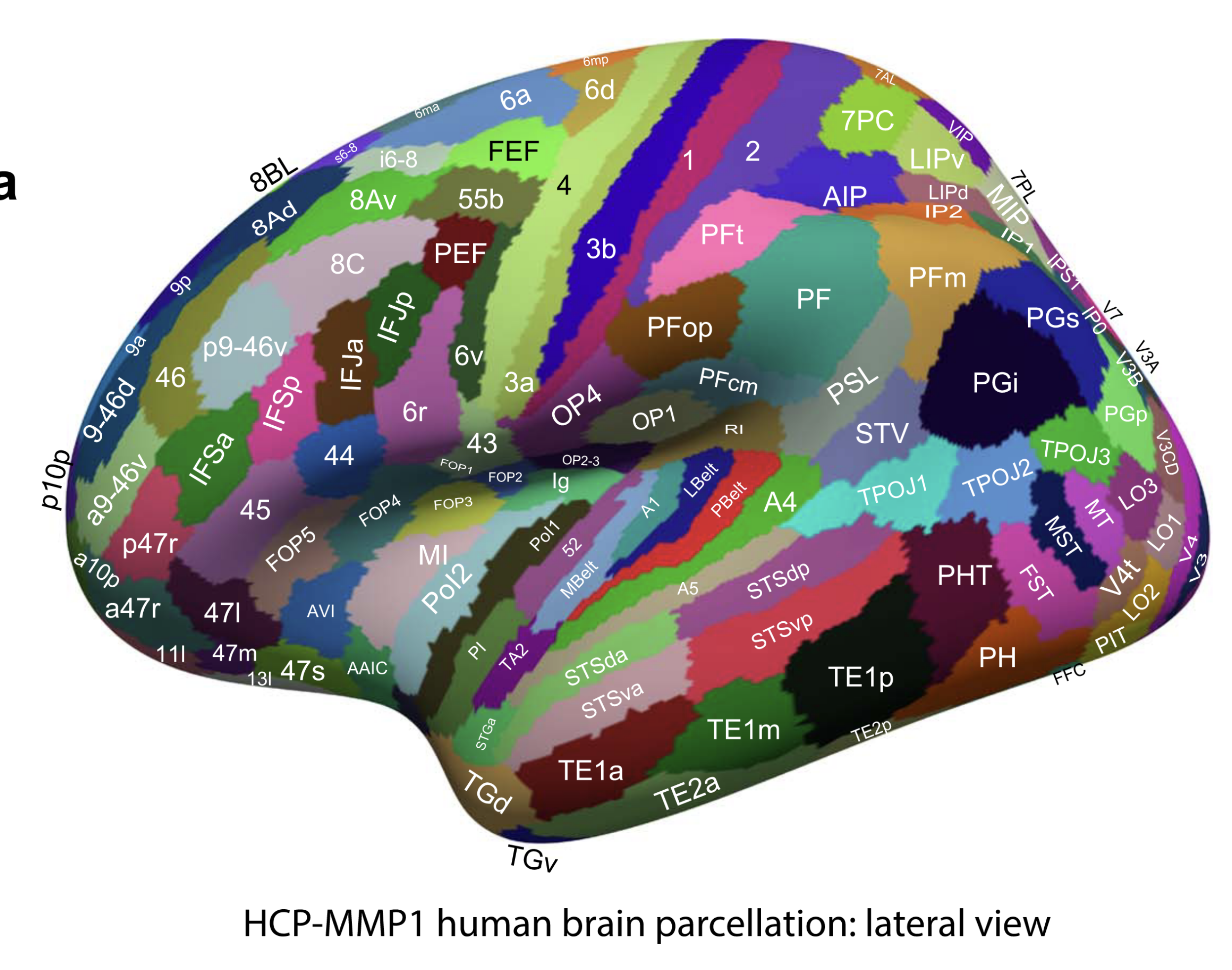
- The early auditory areas include A1, LBelt (Lateral Belt), MBelt (Medial Belt), PBelt (Para-Belt), and the retro-insular cortex (RI). These areas are surrounded by areas, OP2-3, OP1, PFcm, PSL, A4, Ig, and TA2.
- A1 is very heavily myelinated, even relative to its heavily myelinated surrounding neighbors
- interesting colour asymmetries occur in a few areas, especially language-related areas 55b, PSL, SFL, and 44 and their right hemisphere homologues, which also have asymmetric task-fMRI functional profiles
⇒ jetzt verstehe ich auch, warum die Belt regions so langgezogen sind. Sie umgeben einfach den A1 - das macht sie so langgezogen uns der ganze bereich hat eben sehr ähnliche connectivity.
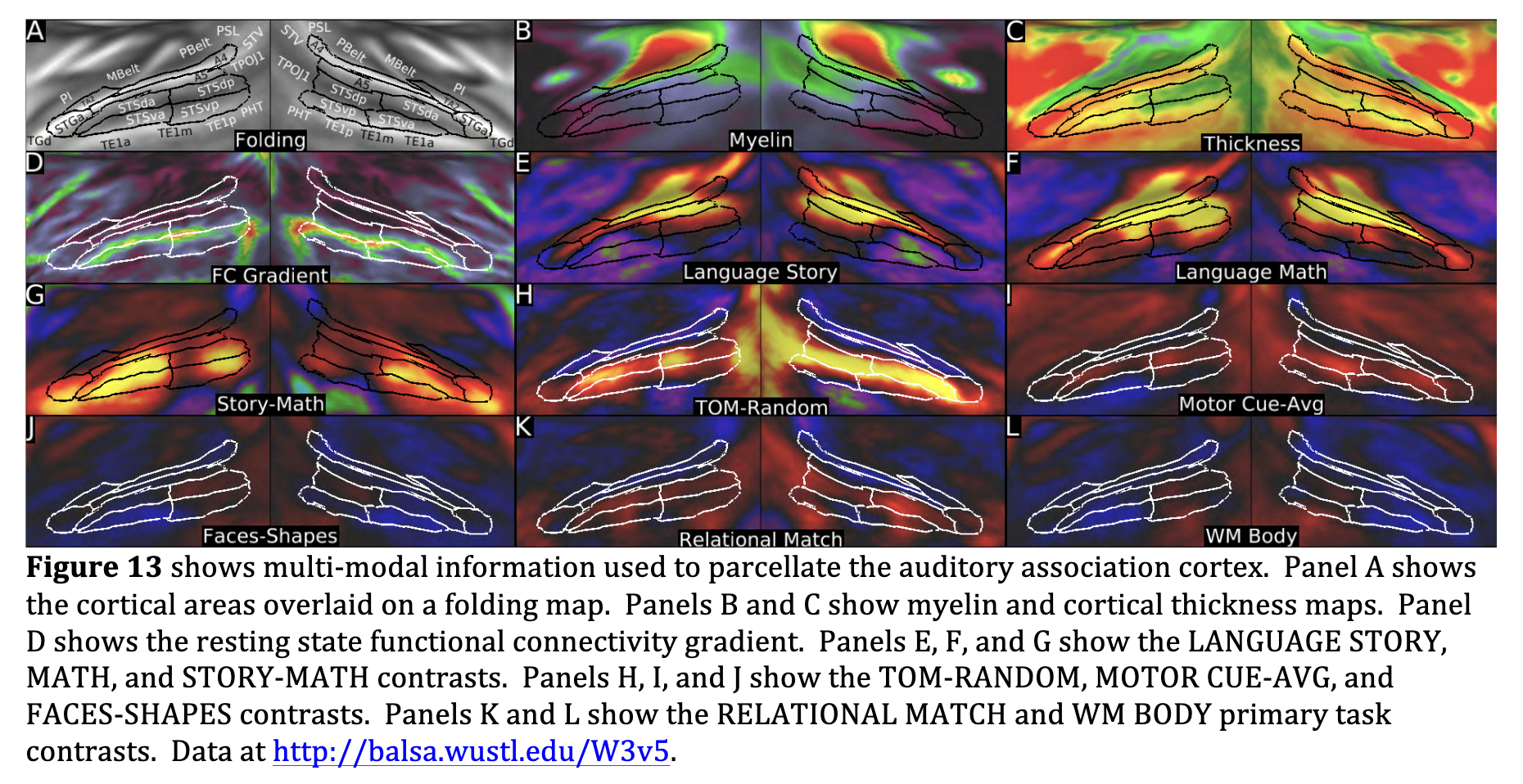
- Relative to its antero-medial neighbor area 52, the MBelt complex has more myelin (Panel B), is thicker (Panel D), and is activated vs deactivated in the LANGUAGE MATH and STORY contrasts (Panel J). Relative to its antero-lateral neighbor area TA2, MBelt has more myelin (Panel B), is thinner (Panel D), and is more activated in the LANGUAGE MATH and STORY contrasts (Panel J). Relative to its lateral neighbor PBelt, the MBelt complex has more myelin (Panel C) and is less activated in the language MATH and STORY contrasts (Panel J) and more activated in the EMOTION FACES-SHAPES contrast.
- We identified auditory association cortex as a region mainly on the superior temporal gyrus and within the superior temporal sulcus that is activated in the LANGUAGE STORY, MATH, and STORY-MATH contrasts.
- It is strongly functionally connected with the inferior frontal gyrus, including areas 44, 45, and 47l.
- This auditory region likely becomes progressively less purely auditory and more multi-modal as one progresses inferiorly, anteriorly, and posteriorly (away from early auditory cortex, e.g. Main Text Figure 3).
- A4, A5, STSdp, STSda, STSvp, STSva, STGa, and TA2
- we have introduced largely novel terminology here, except that TA2 is based on the Von Economo and Koskinas parcellation (Triarhou, 2007a, b; von Economo and Koskinas, 1925). These areas are surrounded by PBelt, MBelt, PI, TGd, TE1a, TE1m, TE1p, PHT, TPOJ1, STV, and PSL.
more about A4:
A4’s supero-medial border with PBelt was covered in Section #10 Early Auditory Cortex. Relative to its inferior neighbor A5, area A4 differs in functional connectivity, and this gradient was primarily used to define the boundary (Panel D). A4 also has more myelin than A5 assessed statistically (Panel B), though the myelin gradient peak does not align with the functional connectivity gradient peak. Relative to its antero-medial neighbor TA2, area A4 has more myelin (Panel B), differs in functional connectivity (Panel D), is more activated in the LANGUAGE MATH and STORY contrasts (Panels E and F), and is less activated in the CUE-AVG contrast (Panel I).
more about A5
- A5 differs in functional connectivity (Panel D), and is more activated in the LANGAUGE STORY-MATH (Panel G) and TOM-RANDOM (Panel H) contrasts. Relative to its inferior neighbor STSdp, area A5 differs in many primary and non-primary task contrasts including more activation in the LANGAUGE MATH (Panel E) and STORY (Panel F) contrasts and less activation in the working memory (e.g Panel L) and RELATIONAL (e.g. Panel J) primary contrasts, and the FACE-AVG, TOM-RANDOM (Panel H), and FACES-SHAPES (Panel K) contrasts. Relative to its inferior neighbor STSda, area A5 again shows differences in a variety of task contrasts including markedly more activation in the LANGAUGE MATH contrast (Panel E), more activation in the TOOL-AVG contrast, markedly less activation in the TOM-RANDOM contrast (Panel H), and less activation in the CUE-AVG (Panel I) and FACES-SHAPES (Panel K) contrasts. Relative to its anterior neighbor STGa, area A5 has more myelin (Panel B) and differs in functional connectivity (Panel D).
more about STSdp:
- Relative to area STSvp on the inferior bank of the STS, area STSdp on the superior banks is has more myelin (Panel B), differs markedly in functional connectivity (Panel D), is more activated in the LANGUAGE MATH (Panel E), TOM-RANDOM (Panel H, especially in the right hemisphere), and MOTOR CUE-AVG (Panel I) contrasts. Relative to its anterior neighbor STSda in the superior bank of the STS, area STSdp has more myelin (Panel B), and differs markedly in its functional activation profile, being more activated in the CUE-AVG contrast (Panel I) and the RELATIONAL MATCH (Panel J), working memory (e.g. Panel L), SOCIAL TOM and other primary contrasts, and less active in the STORY-MATH contrast (Panel G).
⇒ Was bedeuten die Vergleiche aus Glasser SUPPL
see also
Tags: neuroscience science source
Superlink: 050 🧠Neuroscience
Created: 2025-11-12 22:14